Which Labs Read the Results of the for Pemphigus Vulgaris
Pemphigus vulgaris is an uncommon, potentially fatal, autoimmune disorder characterized by intraepidermal blisters and extensive erosions on patently salubrious skin and mucous membranes. Diagnosis is by skin biopsy with directly and indirect immunofluorescence and enzyme-linked immunosorbent analysis (ELISA) testing. Handling is with corticosteroids and sometimes other immunosuppressive therapies.
Bullae are elevated, fluid-filled blisters ≥ 10 mm in diameter.
Skin cleavage levels in pemphigus and bullous pemphigoid
Pemphigus foliaceus blisters form in the superficial layers of the epidermis. Pemphigus vulgaris blisters can class at any epidermal level but typically form in the lower aspects of the epidermis. Bullous pemphigoid blisters form subepidermally (lamina lucida of the basement membrane zone). In this effigy, the basement membrane zone is disproportionately enlarged to display its layers.
Pemphigus (vulgaris, foliaceus, or both) may coexist with certain central nervous system (CNS) disorders, especially dementia, epilepsy, and Parkinson disease. Desmoglein 1 is nowadays in CNS neurons (and in all epithelial cells), and an immunologic cross-reaction betwixt epithelial and CNS isoforms has been suggested.
-
1. Russo I, De Siena FP, Medico, Saponeri A, et al: Evaluation of anti-desmoglein-ane and anti-desmoglein-3 autoantibody titers in pemphigus patients at the time of the initial diagnosis and after clinical remission. Medicine (Baltimore) 96(46):e8801, 2017. doi: ten.1097/Doctor.0000000000008801
Symptoms and Signs of Pemphigus Vulgaris
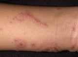
Flaccid bullae, which are the primary lesions of pemphigus vulgaris, cause widespread and painful skin, oral, and other mucosal erosions. Almost one-half of patients have just oral erosions, which rupture and remain every bit chronic, painful lesions for variable periods. Often, oral lesions precede skin involvement. Dysphagia and poor oral intake are common considering lesions also may occur in the upper esophagus. Cutaneous bullae typically arise in normal-appearing skin, rupture, and exit a raw expanse with crusting. Itching is usually absent. Erosions often go infected. If big portions of the trunk are afflicted, fluid and electrolyte loss may be significant.
A rare variant called pemphigus vegetans occurs primarily in intertriginous areas and oral mucosa with vegetating cauliflower-like plaques.
-
Biopsy with immunofluorescence testing
Pemphigus vulgaris should be suspected in patients with unexplained chronic mucosal ulceration, especially if they have bullous skin lesions. This disorder must be differentiated from other disorders that cause chronic oral ulcers and from other bullous dermatoses (eg, pemphigus foliaceus Pemphigus Foliaceus Pemphigus foliaceus is an autoimmune blistering disorder in which lack of adhesion in the superficial epidermis result in cutaneous erosions. Diagnosis is by skin biopsy and straight immunofluorescence... read more 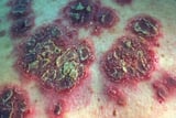 , bullous pemphigoid Bullous Pemphigoid Bullous pemphigoid is a chronic autoimmune skin disorder resulting in generalized, pruritic, bullous lesions in older patients. Mucous membrane involvement is rare. Diagnosis is by peel biopsy... read more
  , mucous membrane pemphigoid Mucous Membrane Pemphigoid Mucous membrane pemphigoid is the designation given to a heterogeneous group of rare chronic autoimmune disorders that tend to crusade waxing and waning bullous lesions of the mucous membranes... read more than , drug eruptions Drug Eruptions and Reactions Drugs tin cause multiple skin eruptions and reactions. The most serious of these are discussed elsewhere in THE MANUAL and include Stevens-Johnson syndrome and toxic epidermal necrolysis, hypersensitivity... read more than
 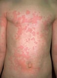 , toxic epidermal necrolysis Stevens-Johnson Syndrome (SJS) and Toxic Epidermal Necrolysis (TEN) Stevens-Johnson syndrome and toxic epidermal necrolysis are severe cutaneous hypersensitivity reactions. Drugs, especially sulfa drugs, antiseizure drugs, and antibiotics, are the near common... read more
 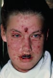 , erythema multiforme Erythema Multiforme Erythema multiforme is an inflammatory reaction, characterized by target or iris skin lesions. Oral mucosa may exist involved. Diagnosis is clinical. Lesions spontaneously resolve but oftentimes... read more
 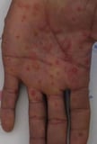 , dermatitis herpetiformis Dermatitis Herpetiformis Dermatitis herpetiformis is an intensely pruritic, chronic, autoimmune, papulovesicular cutaneous eruption strongly associated with celiac disease. Typical findings are clusters of intensely... read more
  , bullous contact dermatitis Contact Dermatitis Contact dermatitis is inflammation of the skin caused by direct contact with irritants (irritant contact dermatitis) or allergens (allergic contact dermatitis). Symptoms include pruritus and... read more
  ).
).
2 clinical findings, both reflecting lack of epidermal cohesion, that are somewhat specific for pemphigus vulgaris are the following:
-
Nikolsky sign: Upper layers of epidermis move laterally with slight pressure or rubbing of peel adjacent to a bulla.
-
Asboe-Hansen sign: Gentle pressure level on intact bullae causes fluid to spread away from the site of pressure and beneath the adjacent pare.
The diagnosis of pemphigus vulgaris is confirmed by biopsy of lesional and surrounding (perilesional) normal skin. Immunofluorescence testing shows IgG autoantibodies against the keratinocyte's cell surface. Serum autoantibodies to desmoglein 1 and desmoglein 3 transmembrane glycoproteins can be identified via straight immunofluorescence, indirect immunofluorescence, and enzyme-linked immunosorbent assay (ELISA).
Without treatment, pemphigus vulgaris is often fatal, usually within 5 years of disease onset. Systemic corticosteroid and immunosuppressive therapy has improved prognosis, but death may withal result from complications of therapy.
-
Corticosteroids, oral or Four
-
Sometimes immunosuppressants
-
Sometimes plasma exchange or 4 immune globulin (IVIG)
Handling of pemphigus vulgaris is aimed at decreasing production of pathogenic autoantibodies. The mainstay of handling is systemic corticosteroids. Some patients with few lesions may respond to oral prednisone 20 to xxx mg once a day, but most require 1 mg/kg once a twenty-four hours as an initial dose. Some clinicians begin with fifty-fifty higher doses, which may slightly hasten initial response just do not appear to better effect. If new lesions go along to appear afterwards 5 to 7 days, Four pulse therapy with methylprednisolone one g once a twenty-four hour period can be tried.
Once no new lesions have appeared for seven to 10 days, corticosteroid dose should be tapered monthly by virtually x mg/mean solar day (tapering continues more slowly once 20 mg/solar day is reached). A relapse requires a return to the starting dose. If the patient has been stable after a year, a trial without treatment can exist attempted simply must be closely monitored.
Immunosuppressants such as rituximab (1 Treatment references Pemphigus vulgaris is an uncommon, potentially fatal, autoimmune disorder characterized by intraepidermal blisters and extensive erosions on plain good for you skin and mucous membranes. Diagnosis... read more 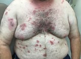 ), methotrexate, cyclophosphamide, azathioprine, gold, mycophenolate mofetil, or cyclosporine can reduce the need for corticosteroids and thus minimize the undesirable effects of long-term corticosteroid use. Rituximab combined with systemic corticosteroids is an culling kickoff-line treatment (two Treatment references Pemphigus vulgaris is an uncommon, potentially fatal, autoimmune disorder characterized by intraepidermal blisters and extensive erosions on apparently salubrious pare and mucous membranes. Diagnosis... read more than
  ). Plasma exchange and loftier-dose IV allowed globulin to reduce antibody titers have too been effective.
-
1. Craythorne EE, Mufti G, DuVivier AW: Rituximab used as a kickoff-line single amanuensis in the treatment of pemphigus vulgaris. J Am Acad Dermatol 65(5):1064–1065, 2011. doi: x.1016/j.jaad.2010.06.033
-
ii. Joly P, Maho-Vaillant Grand, Prost-Squarcioni C, et al: First-line rituximab combined with short-term prednisone versus prednisone solitary for the handling of pemphigus (Ritux three): A prospective, multicentre, parallel-grouping, open-label randomised trial. Lancet 389(10083):2031–2040, 2017. doi: 10.1016/S0140-6736(17)30070-three
-
Nearly half of patients with pemphigus vulgaris accept merely oral lesions.
-
Apply Nikolsky and Asboe-Hansen signs to help clinically differentiate pemphigus vulgaris from other bullous disorders.
-
Confirm the diagnosis past immunofluorescence testing of skin samples.
-
Care for with systemic corticosteroids, with or without other immunosuppressive therapies (drugs, Iv allowed globulin, or plasma exchange).
Source: https://www.msdmanuals.com/professional/dermatologic-disorders/bullous-diseases/pemphigus-vulgaris
0 Response to "Which Labs Read the Results of the for Pemphigus Vulgaris"
Post a Comment